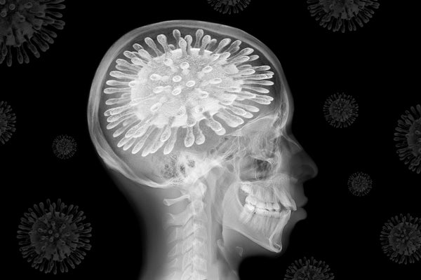
“Brain fog” is not a formal medical descriptor. But it aptly describes an inability to think clearly that can turn up in multiple sclerosis, cancer or chronic fatigue. Recently, the condition has grabbed headlines because of reports that it afflicts those recovering from COVID-19.
COVID’s brain-related symptoms go beyond mere mental fuzziness. They range across a spectrum that encompasses headaches, anxiety, depression, hallucinations and vivid dreams, not to mention well-known smell and taste anomalies. Strokes and seizures are also on the list. One study showed that more than 80 percent of COVID patients encountered neurological complications.
The mystery of how the virus enters and then inhabits the brain’s protected no-fly zone is under intensive investigation. At the 50th annual meeting of the Society for Neuroscience, or SFN (held in virtual form this month after a pandemic hiatus in 2020), a set of yet-to-be-published research reports chronicle aspects of the COVID-causing SARS-COV-2 virus’s full trek in the brain—from cell penetration, to dispersion among brain regions, to disruption of neural functioning.
On supporting science journalism
If you're enjoying this article, consider supporting our award-winning journalism by subscribing. By purchasing a subscription you are helping to ensure the future of impactful stories about the discoveries and ideas shaping our world today.
Trying to find the virus’s port of entry into nerve cells has perplexed investigators, because the surfaces of these cells appear to lack the molecular anchor points—the ones found in lung cells, for instance—that are needed for a forced invasion into the cell interior. Another possible means of ingress was flagged in a study published in Science last year. It showed that the receptor NRP1, present on nerve cells both in the brain and in the olfactory tract, couples with an enzyme on the surface of these cells called furin that permits viral passage.
Still, the question remained: Is this a preferred route into a cell? At an SFN 2021 press briefing, researchers from the All India Institute of Medical Sciences-Patna reported on performing a computer analysis of gene and protein data that showed the presence of NRP1 and furin on cells in some brain areas—particularly the hippocampus, the major memory and learning locus.
Another portal may be situated in the peripheral nervous system, which conveys sensory and motor impulses from muscles, organs and skin to the brain and spinal cord. Jonathan Joyce, a doctoral student in the lab of Andrea Bertke Virginia Polytechnic Institute and State University, explained how his research team infected mice with the SARS-COV-2 virus—and then located viral RNA (instructions for making proteins) as well as viral proteins and the virus itself. They were lodged in clusters of peripheral nerves that had not been previously considered possible entry points. From these nerve groupings, connections stretched out to various areas of the brain. “These routes may be used by SARS-COV-2 to invade the brain,” Joyce says, adding that they may also help explain the nerve pain and tingling that some COVID patients experience.
A consensus has by no means emerged about what exactly occurs during a viral invasion of the brain. Walter J. Koroshetz, director of the National Institute of Neurological Disorders and Stroke, said during a separate SFN 2021 press event that definitive evidence of SARS-COV-2 infecting neurons is “controversial.”
“As an NIH director, I would probably punt and say we’ll have to see how the evidence comes out in the end,” Koroshetz said. Other researchers have suggested that COVID neurological symptoms might be caused by inflammation, by leakage of the blood brain barrier, or by mucosal cells in the lining of the nose becoming infected and dying, leading to the death of nearby neurons as well.
Still another question that captures attention of some labs is where the virus goes once it enters the brain. John H. Morrison, professor of neurology at the University of California Davis School of Medicine and director of the primate research center located there, described his group’s work in looking at viral dispersal. In their study, rhesus monkeys—including a subgroup with diabetes—were infected with SARS-CoV-2. After a week the researchers found traces of the virus (proteins and genetic material) that had spread across several regions of the cerebral cortex, especially in the diabetic animals. The team also discovered that inflammation in the olfactory cortex occurred at the same time as the death of neurons. “The direct entry [of the virus] to the olfactory system, productive infections of neurons, and transport to multiple brain regions are likely the cause of neurological complications in COVID-19,” Morrison says. One area where the virus was present in the diabetic monkeys was the entorhinal cortex, what Morrison calls the “single most vulnerable brain region to Alzheimer’s disease. So once the virus is there, it can play into mild cognitive impairment and dementia.”
One further line of inquiry pursues the impact COVID has had on brain functioning. Electroencephalography measurements conducted by several Canadian institutions—the Rotman Research Institute, McMaster University, the University of Toronto and the Sunnybrook Institute—revealed that even mild COVID cases can lead to altered brain activity. The researchers looked at 42 individuals who had tested positive and then quarantined at home, and compared them with 14 others who had flu-like symptoms but whose COVID test results were negative.
A composite measure of brainwave strength was lower in the COVID group than in the control participants when assessed, on average, four months later. Some of the aberrant signaling had resolved at eight months in those who had tested positive for COVID. But that group as a whole still had lower readings for some measurements; the combination of COVID and social distancing may have had lingering consequences. “The direct effect COVID seems to have on EEG power is analogous to effects that we see when people are diagnosed with mild cognitive impairment, which can develop into Alzheimer’s disease and related dementias,” says Allison B. Sekuler, senior scientist at the Rotman Research Institute. “Now, that doesn’t necessarily mean that everyone who has COVID will end up with Alzheimer’s. But it certainly does warrant more study to determine if the direct effects of COVID on the brain also increase dementia risk.”
The various findings trace how the virus journeys into the brain, but also leave a set of unresolved issues. Rita Balice-Gordon, who moderated and helped organize one of the SFN press briefing but was not involved with research, says the work “demonstrates the tremendous advances that have been made over the last 20 months in understanding the way this virus affects the central nervous system.” She added that it also highlights many questions that remain to be answered such as how long infections persist, how long the neurological and psychiatric symptoms of COVID last and whether the damage caused may lead to a higher risk of dementia and other complications. Balice-Gordon is chief executive of Muna Therapeutics, which develops therapies
The pandemic raises the prospect of growing collaborations between virologists and neuroscientists. It is a reminder that the brain, notwithstanding the blood-brain barrier, is by no means impenetrable. Coxsackie virus, polio, varicella, Epstein–Barr and adenoviruses, among others, can get into brain cells; adenoviruses are even used to deliver gene therapies within the organ. Understanding the distinctive ways that viruses can get in and wreak havoc may draw growing interest. “This is a big issue,” Morrison says. “And I actually think this is going to become a major, major area of study for Society for Neuroscience members.” Integration of the two disciplines is just beginning. Neurovirology may take a place alongside sessions on long-term memory, synapses and glia at some future SFN meeting.